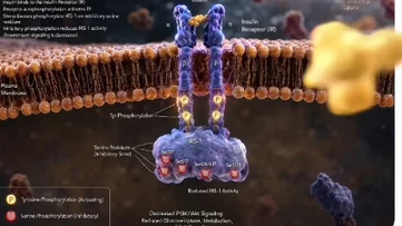
IRS-1 Phosphorylation Inside a Cell
Kling AIPopular cases:Kling AI Video 12。@Image1Create an ultra-realistic, Hollywood-quality 3D biomedical animation of the inhibitory phosphorylation of IRS-…
The idea / Kling AI
IRS-1 Phosphorylation Inside a Cell
Kling AIPopular cases:Kling AI Video 12。@Image1Create an ultra-realistic, Hollywood-quality 3D biomedical animation of the inhibitory phosphorylation of IRS-1 (Insulin Receptor Substrate-1) inside a living human cell. The animati…
Source prompt
@Image1Create an ultra-realistic, Hollywood-quality 3D biomedical animation of the inhibitory phosphorylation of IRS-1 (Insulin Receptor Substrate-1) inside a living human cell. The animation should look like a high-budget scientific documentary from Nature, Cell, or HHMI BioInteractive, with cinematic lighting, ray tracing, shallow depth of field, and photorealistic molecular detail.

Begin with a close-up of a highly detailed phospholipid bilayer plasma membrane, showing the hydrophilic phospholipid heads and hydrophobic fatty acid tails gently moving as in a living cell. Label the extracellular space above the membrane and the cytoplasm below. A realistic insulin receptor spans the membrane as a transmembrane receptor tyrosine kinase. An insulin molecule approaches from the extracellular space and binds to the alpha subunits of the insulin receptor.

Upon binding, the receptor undergoes a realistic conformational change and autophosphorylates on tyrosine residues, shown as glowing golden phosphate groups attaching to the intracellular beta subunits. The camera then zooms toward IRS-1, docked beneath the activated insulin receptor. Initially, IRS-1 appears healthy and ready to transmit insulin signaling. Next, introduce stress kinases such as JNK, IKKβ, mTOR/S6K1, and PKC approaching IRS-1 from different directions.

Each kinase transfers phosphate groups specifically onto serine residues of IRS-1. Each phosphorylation event should produce a brief glow and highlight the modified serine sites. As inhibitory serine phosphorylation accumulates, IRS-1 gradually changes its shape, loses flexibility, and detaches partially from the insulin receptor. Red warning colors appear around the phosphorylated serine residues, indicating impaired function.

Show that downstream insulin signaling is interrupted. The PI3K-Akt signaling pathway fades, with weakened molecular signaling traveling through the cytoplasm. The activation