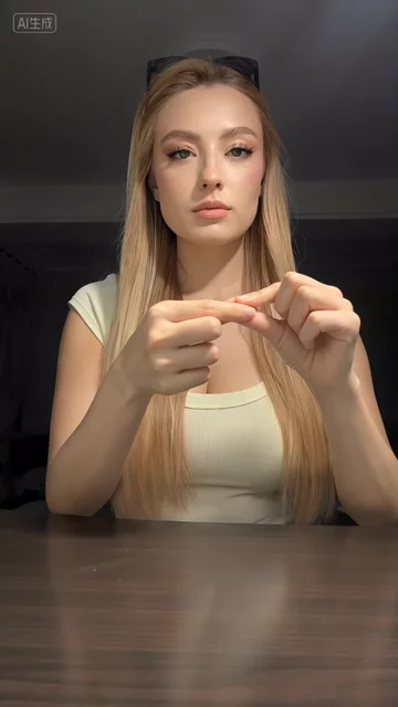
 Colored geometric film between the fingers
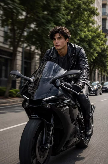 Front tracking on a tree-lined motorcycle ride
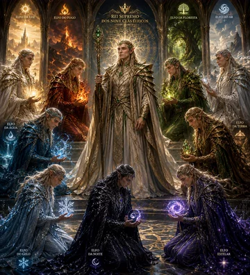 Elven Banquet Animation: AI Scene Prompt
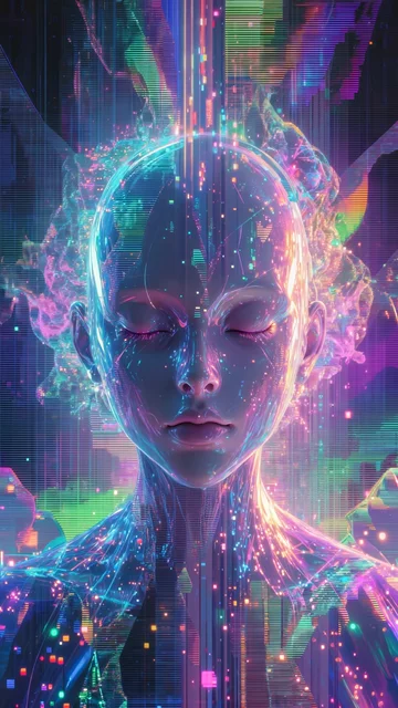 Neon Humanoid Face Slowly Opening Its Eyes
@matkinson
Source promptimage2Subject: a symmetrical synthetic humanoid face made of neon light and digital particles, eyes closedMovement: glitch lines flicker, coloured light streams flow upward, particles converge toward the forehead, subtle breathing motion emerges, facial muscles activate, eyes slowly openScene: abstract digital void filled with vertical data streams and chromatic distortionCamera: slow push-in, perfectly centred, minimal motionLighting: high-contrast neon cyan, magenta, and violet, internal glow from within the faceAtmosphere: surreal, awakening, slightly unsettling, sense of consciousness forming.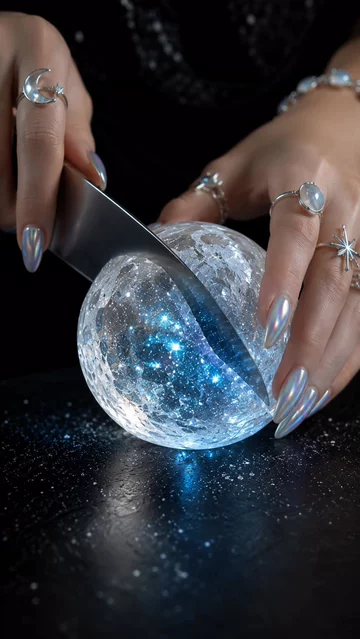 Crystal Moon Cutting ASMR: AI Video Prompt and Breakdown
@janbech
Source promptUltra-realistic macro ASMR video of elegant feminine hands gently holding a flawless glowing moon made of translucent crystal on a matte black stone cutting board. The hands have soft fair skin, slender fingers, glossy pearlescent chrome almond-shaped nails that match the moon’s blue and silver glow, delicate silver celestial rings featuring stars, moons, and tiny gemstones, and a thin moonstone bracelet. The jewelry subtly reflects the crystal’s light. A mirror-polished Japanese chef’s knife slowly slices through the glowing moon. The blade glides effortlessly, revealing shimmering layers of sapphire, silver, and tiny galaxies suspended inside the crystal. Fine sparkling crystal dust falls naturally with each perfectly smooth slice. Every movement is slow, graceful, and intentional. Extreme macro photography, shallow depth of field, cinematic studio lighting, hyper-realistic textures, crisp 8K detail, satisfying ASMR pacing, realistic physics, centered composition, black background, seamless looping, vertical 9:16, no text, no watermark. Apple Gorilla Transformation: AI Creature Prompt
 A flower-meadow afternoon and a paper star
 Two Figures Facing Off in a Dark Studio
@gnmQoiyZ
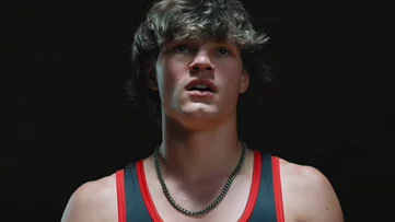
Source promptCLIP 1 — Close-up emoțional (image1):Extreme close-up of image1 looking up slowly with intense emotional expression, mouth slightly open, dark pure black background, single dramatic spotlight, cinematic slow motionCLIP 2 — Reveal (image2):Medium shot of image2 standing alone in dark studio, dramatic spotlight from above, confident pose, slow camera push-in, cinematic tense atmosphereCLIP 3 — Face-off (both):image1 and image2facing each other in pitch black studio, image1 on the left side, image2on the right side, warm amber floor spotlights illuminating each from below, both staring intensely at each other, slow cinematic zoom out, red serif text "YOU" glowing in center between them, dramatic epic atmosphere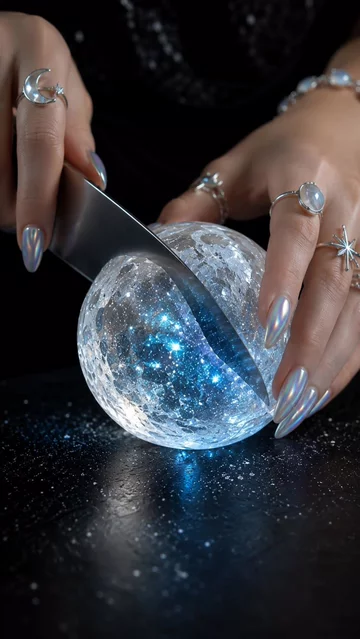 Crystal Moon Cutting ASMR
@not_mac
Source promptUltra-realistic macro ASMR video of elegant feminine hands gently holding a flawless glowing moon made of translucent crystal on a matte black stone cutting board. The hands have soft fair skin, slender fingers, glossy pearlescent chrome almond-shaped nails that match the moon’s blue and silver glow, delicate silver celestial rings featuring stars, moons, and tiny gemstones, and a thin moonstone bracelet. The jewelry subtly reflects the crystal’s light. A mirror-polished Japanese chef’s knife slowly slices through the glowing moon. The blade glides effortlessly, revealing shimmering layers of sapphire, silver, and tiny galaxies suspended inside the crystal. Fine sparkling crystal dust falls naturally with each perfectly smooth slice. Every movement is slow, graceful, and intentional. Extreme macro photography, shallow depth of field, cinematic studio lighting, hyper-realistic textures, crisp 8K detail, satisfying ASMR pacing, realistic physics, centered composition, black background, seamless looping, vertical 9:16, no text, no watermark. A child's delighted reaction to a tablet
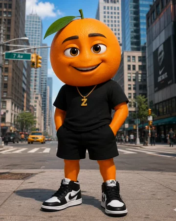 An orange character's street greeting
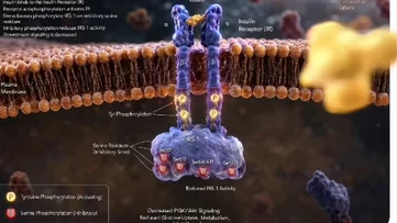 IRS-1 Phosphorylation Inside a Cell
@WjhsykkM
Source prompt@Image1Create an ultra-realistic, Hollywood-quality 3D biomedical animation of the inhibitory phosphorylation of IRS-1 (Insulin Receptor Substrate-1) inside a living human cell. The animation should look like a high-budget scientific documentary from Nature, Cell, or HHMI BioInteractive, with cinematic lighting, ray tracing, shallow depth of field, and photorealistic molecular detail. Begin with a close-up of a highly detailed phospholipid bilayer plasma membrane, showing the hydrophilic phospholipid heads and hydrophobic fatty acid tails gently moving as in a living cell. Label the extracellular space above the membrane and the cytoplasm below. A realistic insulin receptor spans the membrane as a transmembrane receptor tyrosine kinase. An insulin molecule approaches from the extracellular space and binds to the alpha subunits of the insulin receptor. Upon binding, the receptor undergoes a realistic conformational change and autophosphorylates on tyrosine residues, shown as glowing golden phosphate groups attaching to the intracellular beta subunits. The camera then zooms toward IRS-1, docked beneath the activated insulin receptor. Initially, IRS-1 appears healthy and ready to transmit insulin signaling. Next, introduce stress kinases such as JNK, IKKβ, mTOR/S6K1, and PKC approaching IRS-1 from different directions. Each kinase transfers phosphate groups specifically onto serine residues of IRS-1. Each phosphorylation event should produce a brief glow and highlight the modified serine sites. As inhibitory serine phosphorylation accumulates, IRS-1 gradually changes its shape, loses flexibility, and detaches partially from the insulin receptor. Red warning colors appear around the phosphorylated serine residues, indicating impaired function. Show that downstream insulin signaling is interrupted. The PI3K-Akt signaling pathway fades, with weakened molecular signaling traveling through the cytoplasm. The activation